What happens during a lumpectomy and sentinel node biopsy?
Feb 22, 2022*Warning: Graphic content. If surgical pictures are not for you, please skip this post*
You may have a deep desire to help your lumpectomy patients, but not feel confident you actually know what happens in the surgery, which hampers your ability to know how to help your patient during your massage therapy sessions.
If this is you, this digital diary of a lumpectomy + lymph node dissection may be useful, as it highlights from the surgeons point of view what they are doing.
All surgical images are stills from a lumpectomy video listed in JOMI - Journal of Medical Insight (https://jomi.com), which is a peer-reviewed surgical video journal that offers a new gold standard in video-based surgical education.
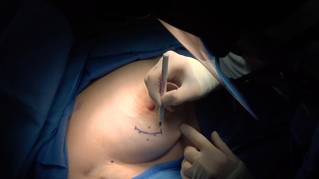
Marking the Patient:
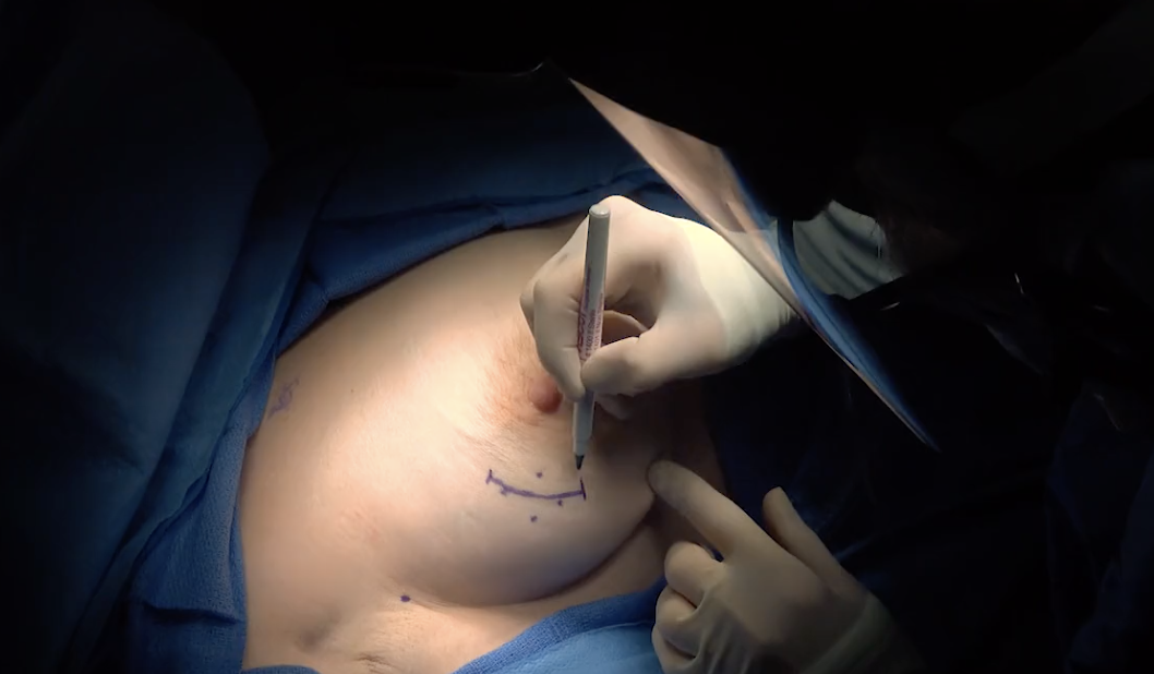 This is done so the surgeon knows where to cut. It is based on both palpation and radioactive signalling, so they know where the tumour and affected lymph nodes are.
Making the incision:
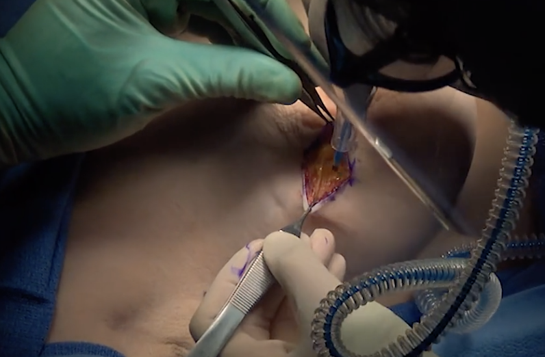 A scalpel is used to split the skin, and then an electrosurgical unit called a "Bovie" is used to incise, destroy tissue by desiccation (dehydration), promote hemostatis (controlled bleeding) and promote blood coagulation.
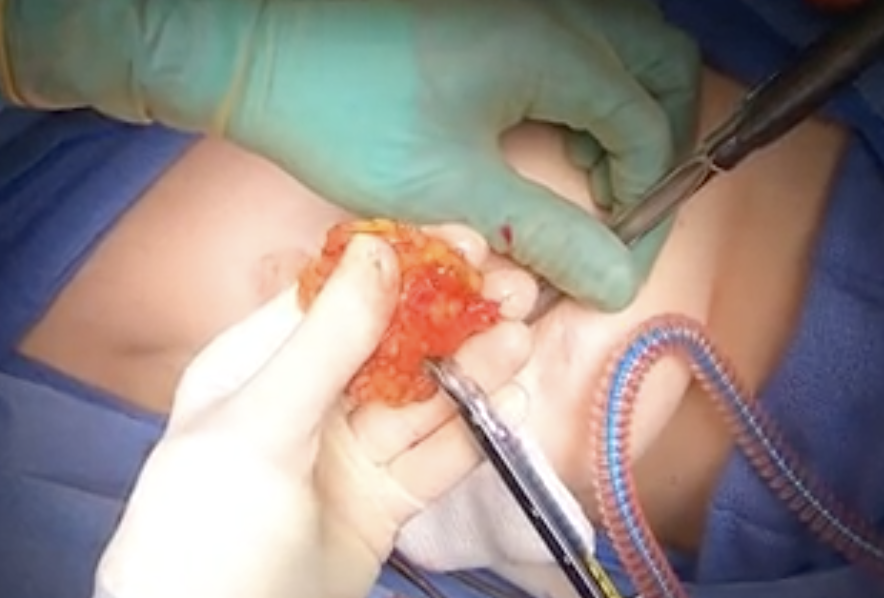
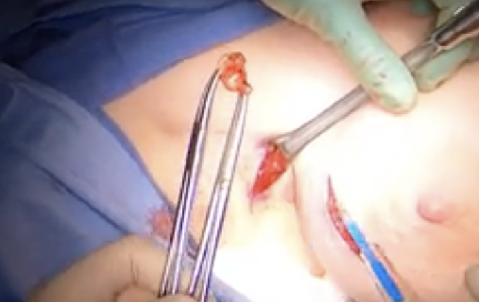
Tumour location & removal:
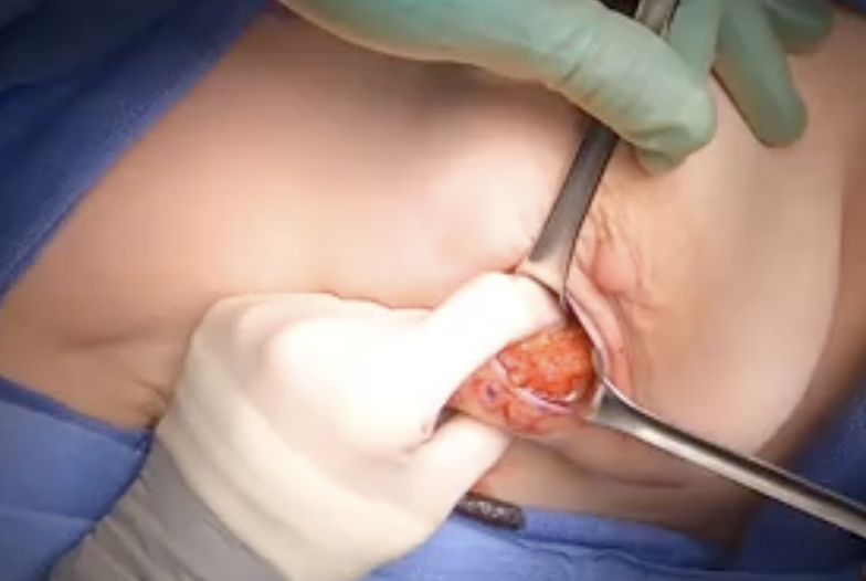
The surgeon hooks her finger around and under the tumour, so she can find the borders and cut away the natural breast fat with the Bovie
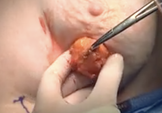
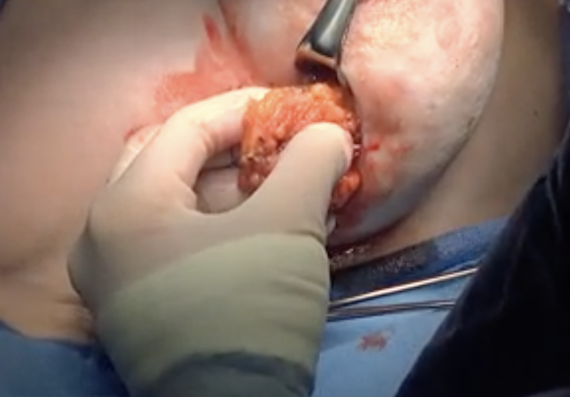
Assessing margins:
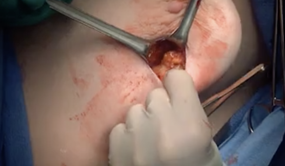
The surgeon in this surgery cut an extra 1.5 cm of breast fat surrounding the tumour, so chances of having clear margins (no cancerous cells) are high.
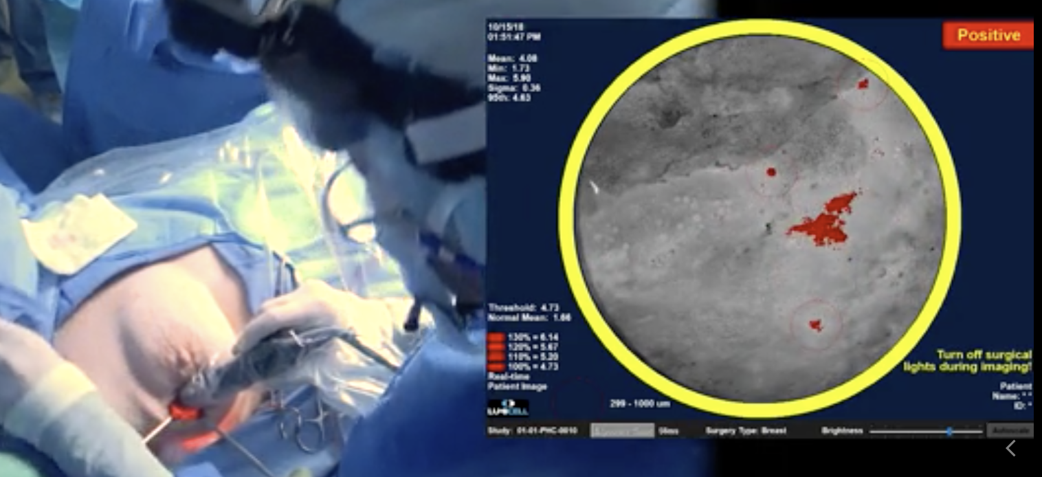
She then continues to asses the surrounding tissues using a Lumicell device to prod all around the excised area for any affected cells remaining. If she finds them, they are then removed.
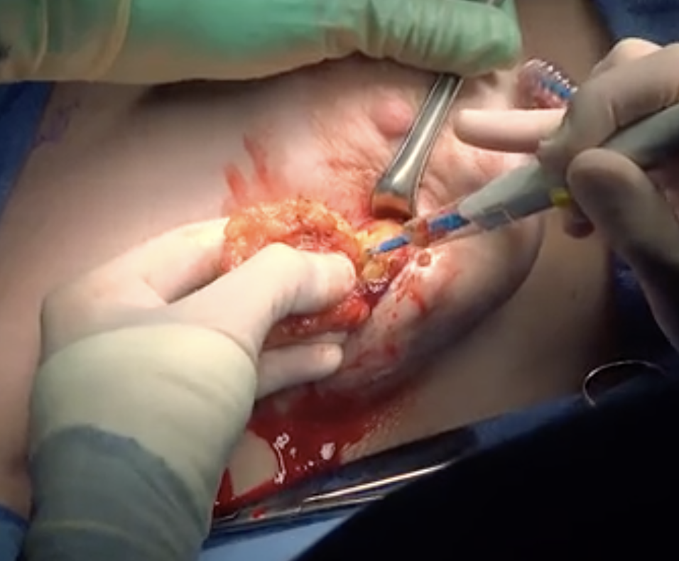
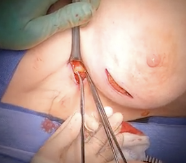
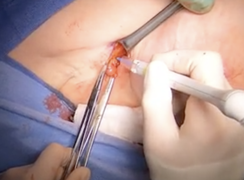
Sentinel node biopsy:

Once the lumpectomy is done and blood vessels cauterized so the wound no longer openly bleeding, the surgeon moves on to the axilla, where she's looking for the lymph node by colour changes done with a radioactive dye, which turns it a mauve colour. She also uses a radioactive probe to test the lymph node and assess whether she has the correct one.
Lymph node location and removal:
Suturing and closing the wounds:
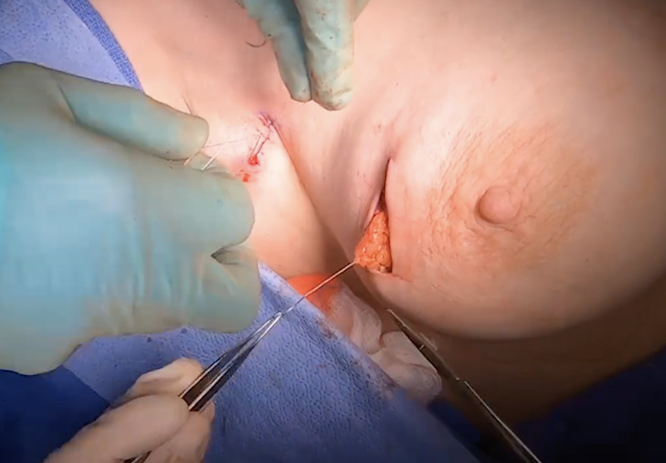
Multiple layers of stitches are done, sewing the breast fat and internal tissues first, before the external stitches are sutured closed.
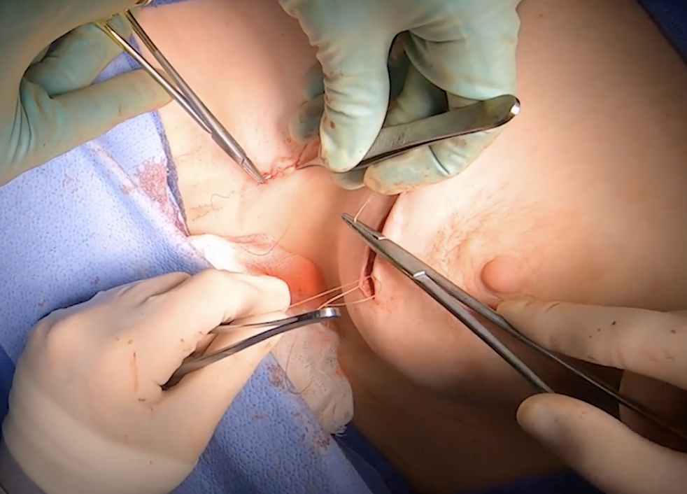
Then the outermost layer of stitches are done with subcuticular stitches so they're not so visible and leave a more pleasing visual after-effect.
So what does this mean for our massage therapy patients?
It means that even though a lumpectomy is a smaller breast-cancer related surgery, you can see just how impactful it is upon the tissues of the breast, axilla, circulatory vessels and lymph pathways. Layers of skin, fascia and fat are cut into, dehydrated, cauterized and removed. Probes go deep and sweep around the interior of the breast cavity.
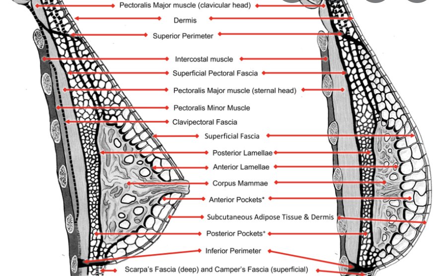
According to Ghaskins, Peoples & McGhee, "The fibro-adipose structure of the female breast is formed by complex scaffolding, consisting of layers of fibrous tissue pockets embedded with adipose tissue which surrounds the corpus mamma and is firmly attached to the perimeter of the breast." (2019).
Think about this image, and the 3D fascial structure of the breast in your treatments, knowing that all layers are interconnected:
Given that the tissues are desiccated and blood vessels are cauterized, it makes sense to me to focus on techniques that rehydrate tissues as well as promote good circulation, with the intention of promoting revascularization and anastomosis.
Fascial techniques to lift, reduce adhesions and restore as much of the 3D structure as possible may be useful, as well as helping multi-layered scar tissue become more flexible and elastic.
In addition, your patient has had a portion of her body removed, which may create a grief and loss response after the surgery, so holding a human-centric healing space for her to recover in may be really useful for her spirits.
If you want to learn more about breast surgery and how you can help your massage therapy patients, please join our Level 1 "Introduction to Breast Surgery" accredited certification course by visiting our Professional Training Program page.
References:
Gaskin, K.M., Peoples, G.E. and McGhee, D.E. (2020), The fibro-adipose structure of the female breast: A dissection study. Clin Anat, 33: 146-155. https://doi.org/10.1002/ca.23505. Retrieved Feb. 22, 2022. Breast image
Jomi. Lumpectomy and Sentinel Lymph Node Biopsy Using Lumicell System for Intraoperative Detection of Residual Cancer. Retrieved Feb. 22, 2022
Science Direct. Electrosurgical Device. Retrieved Feb. 22, 2022
Author